Introduction
There has been renewed interest in mushroom medicinal properties. We studied cholesterol lowering properties of Ganoderma lucidum (Gl), a renowned medicinal species.
Results
Organic fractions containing oxygenated lanosterol derivatives inhibited cholesterol synthesis in T9A4 hepatocytes. In hamsters, 5% Gl did not effect LDL; but decreased total cholesterol (TC) 9.8%, and HDL 11.2%. Gl (2.5 and 5%) had effects on several fecal neutral sterols and bile acids. Both Gl doses reduced hepatic microsomal ex-vivo HMG-CoA reductase activity. In minipigs, 2.5 Gl decreased TC, LDL- and HDL cholesterol 20, 27, and 18%, respectively (P < 0.05); increased fecal cholestanol and coprostanol; and decreased cholate.
Conclusions
Overall, Gl has potential to reduce LDL cholesterol in vivo through various mechanisms. Next steps are to: fully characterize bioactive components in lipid soluble/insoluble fractions; evaluate bioactivity of isolated fractions; and examine human cholesterol lowering properties. Innovative new cholesterol-lowering foods and medicines containing Gl are envisioned.
In Kampo Chinese folk medicine, mushrooms have been known to have medicinal properties since AD1200 [1].
In recent years, there has been interest in the cholesterol lowering properties of mushrooms, including Ganoderma lucidum (Reishi-, Longevity-, or Phantom mushrooms, Biladi Top, Young-zhi, The King Of Herbs, Ling Zhi in Chinese, Saru-no-koshikake and Mannendake in Japanese) [2,3], Pleurotus ostreatus (Oyster mushroom) [4-8], Volvariella volvacea (Straw mushroom) [9], Agaricus bisporus (champignon) [10], Agaricus campestris [11], Auricularia auricula (Tree-ear), Tremella fuciformis (White-jelly leaf) [12,13], Grifola frondosa (Maitake mushroom) [14,15], Lentinus erodes (Shiitake) and isolated fractions [14,16], and Polyporus confluens (Ningyotake) [17]. In an earlier work, Kaneda and Tokuda [18] studied cholesterol lowering properties of ether-, water- and ethanol extracts from caps and stems from Lentinus edodes, Auricularia polytricha (Jews-ear), Flammulina velutipes, and Agaricus bisporus. The majority of these studies were performed in rats. The cholesterol lowering properties of Cordyceps sinensis were studied in humans [19].
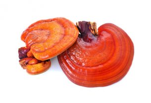
Our focus is Gl, an important medicinal fungus belonging to the Ganodermataceae family that has been studied for its many interesting health promoting properties, including anti-tumor, anti-inflammatory, and anti-platelet aggregation [20-27]http://kyotan.com/lectures/lectures. Indeed, entire books, symposiums, organizations (e.g., the Ganoderma International Research Institute, New York) and therapies have been devoted to Gl. As further testament to its importance, in ancient Chinese times, a Reishi Goddess (Reishi senshi) was even worshipped to bestow health, life and eternal youth.
As described, Gl has been occasionally studied for its cholesterol lowering- and hypotensive properties in the rat [2] and rabbit [28], but not in more physiological cholesterol models [29] such as minipigs. Gl can supposedly lower cholesterol in humans, but the work was not peer-reviewed nor adequately described [24]http://www.ganotherapyusa.com/DXN/docs/whatis.htm.
Like humans, minipigs are omnivours, and their lipid and steroid metabolism, and digestive and cardiovascular physiology closely resembles that of humans [30-32]; whereas in contrast to humans, rodents carry most of the cholesterol in HDL fractions unless they are fed high saturated fat and cholesterol rich diets, which has the effect of shutting down LDL receptors [29].
The components in Gl that may lower cholesterol are not known, but may include ganoderan-type glucans [22,33,34], hetero-β-glucans, glucan-protein complexes (xyloglucans, uronic acid-β-glucans), other fibers, lectins [25], terpenoid triterpenes [35-38], ergostane sterols [39], and highly oxygenated ganoderic acid-type, lanostanoid triterpenes [38-42]. Gl fibrous components could affect cholesterol absorption and bile acid recycling, whereas lipophilic components could affect cholesterol synthesis.
Gl may affect cholesterol synthesis at the committed 3-hydroxy-3-methylglutaryl coenzyme A reductase (HMG-CoA reductase) rate-limiting step; or at the latter lanosterol 14α-methyl demethylase:cytochrome P-450 demethylase (P-45014DM) step [43,44], catalyzing the rate limiting step in lanosterol-cholesterol conversion. In non-Gl mushroom species, inhibition of squalene synthetase by zaragozic acid fungal metabolites has also been reported in primates [45].
Herein, we tested the effects of Gl on cholesterol metabolism in hepatic T9A4 human cells, a hamster small animal model, and a minipig larger animal model having different lipoprotein cholesterol distribution than the hamster model. Animal models were fed cholesterol-containing diets described in Tables Tables1,1, ,22.
Table 1
Proximate analysis of Nafag 924 test diets for hamsters1
| Weight % | |||
| Component | Control | 2.5% Gl | 5.0% Gl |
| Carbohydrate (by difference) | 31.8 | 31.0 | 30.3 |
| Starch | 23.6 | 23.0 | 22.5 |
| Crude Protein | 15.6 | 15.2 | 14.8 |
| Water | 8.0 | 7.8 | 7.6 |
| Crude fat | 4.4 | 4.3 | 4.2 |
| Ash | 4.7 | 4.6 | 4.5 |
| Crude fiber | 3.5 | 3.4 | 3.3 |
| Essential amino acids | 3.4 | 3.3 | 3.2 |
| Gl extract | 0.0 | 2.5 | 5.0 |
| Vitamin mix (includes choline) | 2.4 | 2.3 | 2.3 |
| Minerals (Ca, P, Mg, K, Na) | 2.3 | 2.2 | 2.2 |
| Trace elements | 0.3 | 0.3 | 0.3 |
Table 2
Ingredients in test diets for minipigs1
| Component (g/100 g diet, as fed basis) | Weight % Control or 2.5% Gl |
| Corn (to 100%) | 26.6 |
| Wheat shorts | 29.8 |
| Pork fat2 | 9.0 |
| Soy meal (44%; contains soy protein) | 8.5 |
| Bakery by products | 8.5 |
| Unsalted, melted butter3 | 4.5 |
| Amino acid mix4 | 3.4 |
| Mineral mix5 | 2.8 |
| Cellulose6 (control) or Gl7 | 2.5 |
| Canola (rapeseed) meal | 2.5 |
| Poultry meal | 1.7 |
| Cholesterol8 | 0.1 |
| Vitamin mix (includes choline)9 | 0.1 |
| Trace element mix10 | 0.02 |
Active components in Gl and in vitro activity
Organic and aqueous Gl phases did not contain HPLC-detectable lovastatin. The organic extracted phase strongly inhibited cholesterol biosynthesis (ID50 = 1.3 μg/mL, relative to 0.4 for lovastatin), while the aqueous phase was ineffective (ID50 > 330). Various highly oxygenated lanostanoid triterpenes, and 32-methyl- and 26-oxo sterols were found in the organic phase, and likely contributed to inhibition of cholesterol synthesis. A 20% EtOAc/hexane fraction contained ganoderal A; and a 50% EtOAc/hexane contained ganoderols-A and B, and Y ganoderic acid.
Body and organ weights, and food intake of hamsters
Body weights ranged from 68.7–70.8 and 83.2–86.4 g for the experimental groups on D1 and D18, respectively, without significant differences relative to control, on D1, D18, or D18 minus D1. D18 liver and cecum relative weights (g organ/100 g body wt) were 2.77–2.84 and 0.52–0.56 for the various groups, respectively, without significant differences relative to control. Daily food intake was 7.1–7.8 g food/d averaged over D1-16; there were no significant differences relative to control.
Cholesterol and triacylglycerol in hamsters
Starting D1 TC levels did not differ among the groups, whereas there were differences in D1 TAG (Table (Table3).3). Gl at 2.5 and 5.0% reduced D18 TAG (likely due to D1 TAG differential starting values). Gl at 2.5% did not reduce D18 TC, LDL or HDL. With 5.0% Gl, there was a statistical trend (P < 0.10) to reduce TC and HDL; LDL was not affected. Similarly to the higher dose of Gl, lovastatin decreased D18 TC and HDL, but not LDL. LDL/HDL ratio was not statistically significantly different for any dietary treatments relative to control.
Table 3
Plasma cholesterol and triacylglycerol in hamsters treated with G. lucidum and lovastatin (mmol/L)
| Group | TC | TC | TAG | TAG | VLDL | LDL | HDL | LDL/HDL |
| D1 | D18 | D1 | D18 | D18 | D18 | D18 | D18 | |
| Control | 3.57 | 3.48 | 1.10 | 1.08 | 0.22 | 0.51 | 2.75 | 0.19 |
| Lovastatin | 3.39 | 3.16a | 1.07 | 0.98 | 0.18a* | 0.48 | 2.50a | 0.19 |
| Gl (2.5%) | 3.46 | 3.40 | 0.64a | 0.90a | 0.18 | 0.53 | 2.69 | 0.20 |
| Gl (5%) | 3.34 | 3.14a* | 0.89a* | 0.92a | 0.20 | 0.49 | 2.44a* | 0.20 |
Fecal bile acids and neutral sterols in hamsters
Gl (2.5%) increased fecal total bile acids and chenodeoxycholate (Table (Table4).4). Both Gl doses increased coprostanol 3-one, whereas, 5% Gl decreased cholestanol. Lovastatin had no significant effects on bile acids or neutral sterols examined.
Table 4
Fecal bile acids and neutral sterols in hamsters treated with G. lucidum and lovastatin (nmol/g dry feces/d)
| Bile acids | Neutral Sterols | ||||||||||
| Group | C | LC | DC | CDC | UDC | Total BA | COP-ol | COP-3-one | CHOL erol | CHOL anol | Total NS |
| Control | 15.0 | 14.3 | 10.7 | 5.1 | 1.7 | 46.7 | 11.5 | ND | 7.2 | 18.4 | 37.1 |
| Lovastatin | 15.0 | 12.8 | 11.6 | 6.2 | 1.6 | 47.1 | 12.0 | ND | 6.7 | 18.9 | 37.6 |
| Gl (2.5%) | 16.9 | 16.2 | 14.3 | 8.5a | 1.9 | 57.8a | 11.7 | 4.2a | 7.7 | 16.9 | 40.6 |
| Gl (5%) | 12.2 | 13.9 | 12.0 | 6.4 | 1.4 | 45.8 | 12.0 | 7.8a | 7.0 | 14.1a | 41.0 |
Ex vivo hepatic HMG-CoA reductase activity in hamsters
Lovastatin did not affect de-phosphorylated activity, and phosphorylated activity not examined (Table (Table5).5). In absence of NaF (inhibitor of phosphatase) and in presence of 2.5 and 5 % Gl, 3-hydroxy-3-methylglutaryl-CoA reductase activity in hamster hepatic microsomes (pmol/min/g liver) was reduced 2.1- and and 1.5 fold, respectively, relative to the control. In presence of NaF, 2.5% and 5% Gl reduced HMG-CoA reductase 3.5- and 1.9-fold, respectively, relative to control.
Table 5
HMG-CoA reductase activity in hamster hepatic microsomes (pmol [14C]mevalonolactone/min/mg microsomal protein)
| Group | Activity-NaF | Total activity+NaF |
| Control | 12.36 | 9.30 |
| Lovastatin | 13.34 | ND |
| Gl (2.5%) | 5.86a | 2.66a |
| Gl (5%) | 8.08a | 4.86a |
Fractional cholesterol synthesis rate in hamsters
In hamsters, 24 h FSR values (Atom% enrichment D17-18) were 1.68 ± 0.20, 1.91 ± 0.16, 1.75 ± 0.36, and 2.29 ± 0.05 (mean of n = 6, ± 1 SEM) for control, lovastatin, 2.5%-, and 5% Gl, respectively. Values were not statistically significantly different from control.
Body weights of minipigs
Minipig body weights increased equivalently with Gl and lovastatin from 19.0–26.9 kg over D1-28. Similar weights per age were previously reported for experimentally-fed Göttingen minipigs [46].
Cholesterol and triacylglycerol in minipigs
The experimental diet increased TC 27–30% from D1-14 (Table (Table6).6). In the Gl-fed group, TC significantly decreased 12.5% from D14-21, but not further from D21-29; the decrease in TC from D14-29 was 20% (P < 0.01). Lovastatin did not significantly decrease TC during D14-21 (P > 0.13), D21-29, nor D14-29; but TC did decrease >10% in two pigs from D14-21.
Table 6
Plasma cholesterol and triacylglycerol in minipigs treated with G. lucidum and lovastatin (mmol/L)
| Group | TC | TC | TC | TC | TAG | TAG | TAG | TAG | VLDL | VLDL | LDL | LDL | HDL | HDL | LDL/HDL | LDL/HDL |
| D1 | D14 | D21 | D29 | D1 | D14 | D21 | D29 | D14 | D29 | D14 | D29 | D14 | D29 | D14 | D29 | |
| Gl (2.5%) | 2.47a | 3.21bc | 2.81 | 2.58 | 0.53 | 0.57 | 0.80 | 0.69 | 0.07 | 0.09 | 1.45c | 1.08 | 1.69c | 1.42 | 0.88 | 0.79 |
| Lovastatin | 2.36a | 3.00 | 2.44 | 2.81 | 0.50 | 0.60 | 0.59 | 0.71 | 0.10 | 0.09 | 1.40 | 1.29 | 1.50 | 1.43 | 0.95 | 0.91 |
There were no significant differences in TAG and VLDL with Gl or lovastatin (Table (Table6).6). VLDL was however a minor lipoprotein pool. Lovastatin had not significant effects on LDL nor HDL; Gl decreased LDL 26% and HDL 16% (P < 0.01; D14 vs 29). Gl did not affect statistically significantly affect LDL/HDL since both individual parameters decreased from D14-29.
Fecal bile acids and neutral sterols in minipigs
The high cholesterol-fat diet decreased chenodeoxycholate; and increased coprostanol, coprostan 3-one, and cholesterol from D1-14 (P < 0.05 or < 0.10; Table Table7).7). Gl trended to increase cholestanol (D14 vs 29; P < 0.10).
Table 7
Fecal bile acids and neutral sterols in minipigs treated with G. lucidum and lovastatin (nmol/g dry feces)
| Bile acids | Neutral Sterols | ||||||
| Group | NCT | C | CDC | COP-ol | COP-3-one | CHOL erol | CHOL anol |
| All pigs (D1) | 1.98 | 0.86 | 0.66a | 2.41a | 0.10a | 1.73a* | 0.96 |
| All pigs (D14) | 1.98 | 1.33 | 0.16 | 3.75 | 0.17 | 2.96 | 0.91 |
| Gl (D14) | 1.98 | 1.61 | 0.14 | 3.61 | 0.18 | 3.37 | 0.87b* |
| Gl (D29) | 1.98 | 0.81 | 0.16 | 4.44 | 0.15 | 3.15 | 1.22 |
| Lovastatin (D14) | 1.98 | 0.99 | 0.19 | 3.92 | 0.15 | 2.44 | 0.96 |
| Lovastatin (D29) | 1.98 | 1.23 | 0.18 | 3.28 | 0.16 | 2.34 | 1.14 |
ctive components in Gl and in vitro activity
As described, lovastatin was not detected in our Gl mushroom preparations. By contrast, statin-like compounds have been found in oyster mushrooms [47] and Chrysosporium pannorum [48].
We did however detect oxygenated lanosterol molecules such as 32-methyl- and 26-oxo sterols, ganoderols-A and B, Y ganoderic acid, and ganoderals-A and B in the organic layer. The organic layer strongly inhibited cholesterol biosynthesis from acetate. Similar or identical oxygenated lanosteroids had been previously reported in Gl [38–42], and found to inhibit conversion of 24,25-dihydrolanosterol to cholesterol at the lanosterol 14 α-demethylase step [49–51], and also indirectly to inhibit HMG-CoA reductase activity [51]. The fact that the aqueous phase from Gl was ineffective at inhibiting cholesterol synthesis (ID50 > 330) suggests that hydrophilic molecules such as glucans and fibers in Gl do not affect conversion of acetate to cholesterol. Such molecules may however affect cholesterol absorption and bile acid recycling.
Ex vivo hepatic HMG-CoA reductase and fractional cholesterol synthesis rate in hamsters
The observed inhibition of ex-vivo HMG-CoA reductase activity in hamsters treated with Gl has similarly been observed with Gl in rats [51], and with pure lanosterol analogs [44,52]. Our lack of effect with lovastatin (4.3 μmol/kg body wt) contrasts results with the related statin, simvastatin, where 10, 30, and 60 μmol/kg body wt/d increased ex-vivo hepatic HMG-CoA reductase activity 2-, 17-, and 50-fold, respectively [53]. Lovastatin could have different effects on HMG-CoA reductase and other enzymes than simvastatin, and was not however examined in the above study.
Lanosterol analogs such as those found in Gl are known to inhibit translation of HMG-CoA reductase mRNA, and may also accelerate protein degradation [44,52]. Gl may also affect cholesterol biosynthesis at latter biosynthetic steps such as the conversion of lanosterol [51], which could in turn, indirectly inhibit HMG-CoA reductase activity, as reported for statins in minipigs [53]. Indeed, it was reported that repression of the lanosterol 14 α-demethylase step can result in accumulation of 3 β-hydroxy-lanost-8-en-32-al, a known translational downregulator of HMG-CoA reductase [54].
If Gl had direct physical effects on HMG-CoA reductase activity, this implies that even after the 16 h fast employed in hamsters, Gl components were still bound to the enzyme during the assay procedure [55]. After the 16 h fast, lovastatin could have been removed from the enzyme accounting for the lack of observed effects of lovastatin on ex-vivo HMG-CoA reductase activity. Due to removal of the drug, other statins have even been found to increase ex-vivo HMG-CoA reductase activity [56]. Hepatic ex-vivo HMG-CoA reductase activity and whole body cholesterol FSR are entirely different types of measurements. It is not clear why Gl and lovastatin did not influence cholesterol FSR in hamsters. In principle, the low saturated fat-cholesterol condition employed via use of a chow diet, should have led to a high endogenous rate of cholesterol synthesis, one that could be inhibited by Gl and lovastatin. It is conceivable that the Gl and lovastatin became decomposed in the dietary mixture. To test this hypothesis, we re-extracted Gl and lovastatin from stored diets after culmination of the experiments, and found no differences in bioactive components analyzed, compared to the original starting materials (before addition to the diets; data not shown).
Cholesterol and triacylglycerol in hamsters and minipigs
Hamsters were fed a low-cholesterol chow-based diet with no added exogenous cholesterol or saturated fat. Under these conditions, there was not sufficient cholesterol to redistribute cholesterol from the HDL to LDL pool [29]. This is why in hamsters, 5% Gl and lovastatin reduced D18 TC and HDL, but not LDL [57,58].
Using the same types of diet, lovastatin was similarly found to preferentially reduce HDL in hamsters; and only when dietary saturated fat was added, were both LDL and HDL reduced [57].
Another factor contributing to the lack of strong effects in hamsters, and the total lack of effect in minipigs may be that the dose of lovastatin was insufficient. In hamsters, the employed dose of 2 mg lovastatin/100 g diet is ca. 4.3 μmol lovastatin/kg body wt. Himber et al. [57] treated hamsters with 25 μmol lovastatin/kg body wt, which lowered HDL; or 50 μmol, which lowered LDL and HDL [57]. Morand et al. [53] found that 20–200 μmol simvastatin/kg body wt was sufficient to reduce LDL. Ma et al. [59] reduced lipoproteins in hamsters with 100 mg lovastatin/100 g diet. In minipigs, we utilized a dose of 80 mg lovastatin/minipig/d, which may also have been on the low side. A dose of 24–42 mg was sufficient to lower lipoproteins in Hyde Park minipigs [60]. Nevertheless, our particular species, strain, and location of minipigs may have responded less aggressively to lovastatin (M. Huff, Personal Communication, December 2000). In Göttingen minipigs, a dose of 80 mg simvastatin lowered LDL, whereas 240 mg lowered LDL and HDL [53]; simvastatin is likely more effective in minipigs than lovastatin at a similar dietary weight percent [61,62].
The reduction in TAG with Gl was likely due to lower D1 TAG values in the Gl groups relative to control. TAG reductions in hamster models typically occur under conditions of higher saturated fat intake [6,63]. In the only other peer-reviewed study examining cholesterol lowering properties of Gl in a small animal model, 5 dietary wt% dried Reishi mushroom powder was found to decrease TC in SHR rats; effects on VLDL, LDL and HDL were not studied [2]. In minipigs, with the high fat-cholesterol feeding conditions employed, a Gl-induced inhibition of cholesterol synthesis should result in less availability of hepatic cholesterol for lipoprotein synthesis. In turn, this has the potential effect of reducing plasma VLDL cholesterol secretion, reducing LDL direct secretion; and possibly reducing VLDL-LDL conversion [64,65]. In the present work, we did not observe differences in TAG or VLDL in pigs fed either Gl or lovastatin, however this effect could have been missed since the VLDL pool represented only a small lipoprotein pool and/or there was efficient VLDL-LDL conversion. The reductions in both LDL and HDL with Gl is consistent with that seen with higher statin doses [53].
Fecal bile acids and neutral sterols in hamsters and minipigs
In hamsters, Gl increased fecal total bile acids and chenodeoxycholate, whereas both doses, increased coprostanol 3-one; the 5% dose decreased cholestanol for unclear reasons. An increase in fecal chenodeoxycholate likely indicates production or recycling of chenodeoxycholate was enhanced.
Plasma levels of cholestanol are positively associated with cholesterol absorption [66]; whereas decreased fecal cholestanol may indicate plasma cholestanol was increased and cholesterol absorption was enhanced. In minipigs, Gl tended to increase fecal cholestanol, the opposite pattern to that of hamsters fed 5% Gl. Coprostanol and coprostanol 3-one are the bacterial products of cholesterol, which are increased when fecal cholesterol is increased, or when gut flora are altered [67]. Since fecal cholesterol and coprostanol levels were not changed by either dose of Gl, it is not obvious why coprostanol 3-one accumulated.
Bile salts are now known to possess many different functions acting as detergents, activators of protein kinase C and phosphatidylinositol-3 kinase; and being important gene regulators [68,69]. Chenodeoxycholate, deoxycholate, and their glycine and taurine conjugates can lead to farnesoid X receptor/retinoid X receptor (FXR/RXR)-induced activation of intestinal bile acid binding protein transcription (I-BABP), and suppression of CYP7α RNA and protein levels (FXR prevents liver X receptor (LXRα)-induced transactivation of CYP7α). CYP7α regulates the committed step in classical bile acid synthesis. Overall, an increased fecal level of chenodeoxycholate would mean less chenodeoxycholate is available to activate FXR. Less activation of FXR would lead to less bile acid recycling and less inhibition of bile acid synthesis, more hepatic cholesterol converted to bile acids, and a lowering of plasma cholesterol.
Overall, it is likely that fibrous and/or lipophilic sterol-like molecules in Gl altered the absorption and recycling of bile acids and neutral sterols, leading to altered fecal accumulation. Monitoring plasma levels of neutral sterols and bile acids, and quantifying conjugated and de-conjugated bile acids, should help to clarify the potential importance of the observed trends.
Comparing in vitro, ex vivo, and in vivo results
In the present work, the in vitro experiments were performed with fractionated Gl extracts, whereas the ex-vivo and in vivo work utilized intact Gl. Intact Gl contains fibrous components, which may have affected bile acid and neutral sterol absorption and recycling. Fibrous components could also impair the uptake of lipophilic components, such as those inhibiting in vitro cholesterol synthesis. An additional complexity is that lipophilic components such as ergostane sterols [39] could also affect bile acid and neutral sterol levels. Thus, it is difficult to directly compare our in vitro and in vivo results. Feeding fractionated and intact mushrooms should help to unravel the in vivo bioactive components, as has been accomplished for oyster mushrooms
Conclusions and key findings
In summary, GI was found to have cholesterol lowering potential in vitro, ex-vitro, and in two animal models, with some differences between the two animal models. It is possible that oxygenated lanosterol derivatives in Gl (partly characterized in the present work) contributed to this cholesterol lowering by decreasing cholesterol synthesis (changes in in vitro and ex-vivo, but not whole body, cholesterol synthesis were apparent in the present work). Fibrous components and glucans in Gl were likely responsible for the observed alterations in fecal neutral sterols and bile acids in both animal species, ultimately affecting cholesterol absorption and bile acid recycling and contributing to cholesterol lowering. Next steps are to examine the cholesterol lowering properties of various doses of intact and fractionated, chemically characterized, Gl components in a placebo-controlled clinical trial. Animal experimentation should also utilize fractionated materials, and ideally, elucidate mechanisms of action of each bioactive component. Positive cholesterol-lowering results in such studies will pave the way for adding Gl to new cholesterol-lowering foods and medicines, alone, and in combination with other established cholesterol-lowering ingredients and drugs.
Materials and methods
Materials
Gl was from Fermenta SA, Payerne, Switzerland. Mushrooms were cultivated on a defined formula of sawdust, wheat straw and millet grain. Substrate was sterilized at 90°C for 48 h, then incubated with Gl seed material from Mycotec Sàrl (Cernier, Neuchâtel). Cultivation was with controlled temperature, light, humidity and carbon dioxide concentration. Human hepatic T9A4 cells [71] were grown in LCM serum-free media under 3.5% CO2 at 37°C. Lovastatin was purchased as 20 mg Mevacor tablets (MSD Chibropharm GmbH, Haar, Germany). HMG-CoA reductase, DL-3-Glutaryl-3- [14C]-HMG-CoA (2216 MBq/mmol), R-[5-3H] mevalonic acid ammonium salt (1443 MBq/mmol), and [1-14C] acetic acid sodium salt (2070 MBq/mmol) were from Amersham (Upsala, Sweden). α-3-HMG-CoA (cold) and liquid scintillation cocktail were from Sigma (Buchs, Switzerland). LCM cell medium was from Biofluids (Rockville, MD). 5β-cholesteane-3α-ol, 5-α-cholestane and 2,3-nor-5β-cholanicacid-3α,7α,12α-triol were from Steraloids, Inc. (Newport, Rhode Island); other steroid standards were from Sigma, and Calbiochem (La Jolla, California). Methanolic HCl and Sylon HTP were from Supelco (Buchs, Switzerland). The Cobas Bio autosampler was from Hoffmann-La Roche (Basel, Switzerland) and reagents were from Roche Diagnostics (Rotkreuz, Switzerland). Total Cholesterol Kit 352 and Triacylglycerol Kit 336 were from Sigma. Deuterium was from Cambridge Isotope Laboratories (Andover, MA). Zn catalyst was from Biochemical laboratories (University Bloomington, IN). Silica gel thin layer chromatography (TLC) plates were from Merck Eurolab (Dietikon, Switzerland). Coomassie Plus-200 protein assay reagents and bovine serum albumin fraction V were from Pierce (Rockford, Illinois). All other chemicals were from Sigma.
Preparation of Gl for in vitro testing
Fruiting bodies from Gl (20 g) were dried, milled and macerated in 0.4 L MeOH/H2O (4:1, v/v) at room temperature for 3d. The mixture was then filtered, evaporated, re-dissolved in H2O, acidified to pH 3 with 3 M HCl, extracted 3 × with 150 mL ethyl acetate, and the organic phase evaporated under vacuum at 30°C, re-dissolved in 10 mL MeOH, and dried with Na2SO4, for HPLC analyses and in vitro testing.
Chemical analysis of Gl
The presence of lovastatin in Gl was determined by HPLC with a Nucleosil 100-5 C18 column (250 × 4 mm; Macherey-Nagel, Oensingen, Switzerland) and a Lichrospher 100 RP-18 post column (Merck, Glattbrugg, Switzerland). Solvent A was H3PO4/H2O (1:2000, by vol); solvent B was acetonitrile. Separation was initiated with a linear gradient of 95% A, 5% B, reaching 50% A, 50% B in 45 min, 30% A, 70% B in 46 min, 10% A, 90% B in 48 min, and 0% A, 100% B in 50 min; the run was continued isocratically 4 min. Initial conditions were maintained 6 min for re-equilibration; the flow rate was 1 mL/min. The detector was a G1315 A, series 1100 detector (Hewlett Packard, Meyrin, Switzerland); absorbance was measured at 254 nm. After selective extraction and purification with different adsorbents and solvents, ganoderols and ganoderic acids were detected by mass spectroscopy and NMR (details to be published separately).
In vitro activity of Gl extracts
Human hepatic T9A4 cells were grown in LCM serum-free media under 3.5% CO2 at 37°C. Cells were seeded in 24-well plates and at confluence, incubated with 1 mM 14C-acetate (1 mCi/mmol) for 20 h ± mushroom extracts. Lipids were extracted from cells by incubating 2 × with 1.5 mL hexane/isopropanol (3:2, by vol) for 30 min at room temperature. Combined organic extracts were dried under N2, re-dissolved in hexane, and separated by TLC with hexane/diethyl ether/acetic acid (75:25:1, by vol). Cholesterol synthesis was determined by measuring incorporation of 14C from acetate to cholesterol. Radioactivity was assessed with an instant imager and expressed as percent of control.
Administration of Gl and lovastatin to hamsters
Male Golden Syrian hamsters (Harlan, UK), 3–4 wks, 40–60 g, were housed individually in Macrolon Type 3 cages with 12 h alternating periods of light and darkness. During 3 wks preceding treatment, hamsters were fed Nafag 924 hamster complete diet (# 3132/20, Eberle Nafag AG, Gossau, Switzerland; Table Table1).1). Following body weight randomization, groups consisted of 6 hamsters/group receiving either: Nafag diet (control), Nafag mixed with 2 mg lovastatin /100 g diet (powdered in liquid N2); or Nafag mixed with 2.5 or 5.0% dried Gl. Hamsters were fed experimental diets for 17 d. Lovastatin is an inhibitor of HMG-CoA reductase [72], and was used as a positive control. Dietary intake was recorded daily, body weights weekly. Feces were collected on D15-18. Hamsters were injected subcutaneously with 250 μL D2O on D17 and killed under anesthesia with isoflurane on D18. Following a 16 h fast, D1 (0.5 mL) and D18 blood (>3 mL) were obtained from the retro-orbital cavity and cardiac vein, respectively, and transferred to EDTA tubes. Plasma was prepared by centrifugation at 1500 g, 15 min, at 4°C. Plasma, and hepatic and cecum tissues were stored at -80°C. Animal procedures were authorized by Service Vétérinaire du Canton de Vaud, Switzerland, protocol 1247.
Administration of Gl and lovastatin to minipigs
Nine female and one male Göttingen minipig(s) (Jörg Farm in Bern Switzerland; Minipig-Primärzucht, Auswill, Switzerland) aged 6–12 mo (18–20 kg), with white (7) and black (3 minipigs) colorations, were housed in a 30 m2 box with normal light/dark cycle, and kept at room temperature. Females were chosen because they have fewer age-related lipid modifications and higher lipid concentrations than males [73]. One male was accidentally provided in the delivery, however its total cholesterol (TC), lipoproteins, bile acids and neutral sterols were similar to that of other minipigs. Minipigs were randomly distributed by weight into two separately housed groups, marked with a plastic label in the ear, and fed twice daily for 11 d with powdered commercial pig chow (Diet 574, Minipig-Primärzucht). During a subsequent 4 d adaptation period, minipigs were fed an acclimatization mixture of chow and increasing amounts of powdered hypercholesterolemic control diet (custom diet 2604, Kliba, Kaiseraugst, Switzerland; Table Table2)2) from 0% to 100%, in steps of 25%, designed after Burnett et al. [64,74], that was consistent with Göttingen minipig nutritional needs [75]. During the following 2 wks (D15-29), groups were fed control hypercholesterolemic diet pre-mixed with 2.5% Gl extract; or hypercholesterolemic diet plus 80 mg lovastatin/pig/d (in four 20 mg tablets) [53], hand fed to each minipig, mornings, in half an apple. For acclimatization, on D12-14, minipigs received a half apple without lovastatin. The study was blinded in that the diets were coded, and the mushroom extract was referred to as “Nestlé Special Fiber.” Food intake was 3.5% of body wt/d (based on group average wt), readjusted weekly, to provide sufficient, but not excessive, calories [64,65,75]. Diets were distributed at 0700 and 15h00, and spread linearly on a clean cement surface to facilitate individual consummation. Distilled water was provided ad libitum. Toys and human contact were provided to avoid boredom. Fasting 16 h blood samples (10 mL; 20 mL on D29) were collected in EDTA tubes on D1, 15, 22, 28, and 29 from anterior vena cava. Plasma was prepared by centrifugation as described for hamsters, and stored at -80°C. Blood collection began at 0800 following injection of the intra-muscular relaxant Dormicum® (Hoffmann La Roche, Basel, Switzerland), then the tranqulizer Stresnil® (Janssen Pharmaceuticals, Beerse, Belgium). After blood sampling on D1, 15 and 29, minipigs were isolated for 2 h maximum for individual fecal collections. Some minipigs did not defecate during this period, whereas others defecated again following return to their groups. Hence, the morning fecal collection was qualitative. Feces were stored at -40°C under N2. Body weight was recorded weekly, and food intake recorded each morning. Minipigs were donated to the University of Geneva at the study’s conclusion. Animal procedures were authorized by Service Vétérinaire du Canton de Geneve, Switzerland, protocol 1315, authorization 31.1.1014/1719/1.
Cholesterol and triacylglycerol measurements in hamsters and minipigs
Plasma total cholesterol and triacylglycerol were measured using commercial kits and a Roche Cobas Bio autosampler. Plasma lipoproteins were separated by size-exclusion HPLC as previously described [63].
Fractional cholesterol synthesis rate measurements in hamsters and minipigs
Measurements of water- and cholesterol deuterium enrichment were performed with a Finnigan Thermoquest Delta XL plus Isotopic Ratio Mass Spectrometer (Bremen, Germany) as previously described [76,77]. Fractional synthesis rate (FSR) of free cholesterol was calculated from a plasma sample collected 24 h after deuterium oxide subcutaneous injection as follows: FSR (in % pool/d) = 100 × (cholesterol enrichment/(water enrichment × 0.478)). Due to technical reasons, there were insufficient values in the minipig experiments to reach interpretable conclusions.
Fecal bile acids and neutral sterol measurements in hamsters and minipigs
Fecal neutral sterols and bile acids were extracted from lyopholized feces, deconjugated, derivatized with Sylon HTP and analyzed by gas chromatography as previously described with internal standards: 5-α-cholestane for neutral sterols; 2,3-nor-5β-cholanic acid-3α,7α,12α-triol for bile acids [63].
Hepatic ex-vivo HMG-CoA reductase measurements in hamsters
Freshly excised liver (300 mg) was collected after 16 h fast of hamsters, minced with scissors, and homogenized with 0.4 mL buffer (50 mM KH2PO4, 0.1 M sucrose, 50 mM KCl, 50 mM NaCl, 30 mM EDTA, and 2 mM dithiothreitol, ± 50 mM NaF) with a Potter-Elvehjem S homogenizer with 400 rpm/5 strokes, on ice, after Conde et al. [55]. NaF inhibits dephosphorylation of HMG-CoA reductase by inactivating phosphoprotein phosphatases, yielding total phosphorylated HMG-CoA reductase activity. After washing homogenizer with 0.2 mL buffer, homogenate was centrifuged at 10000 g, 15 min, at 4°C. Supernatant was decanted, 0.4 mL cold buffer added, and the tube vortexed and re-centrifuged. Pooled post-mitochondrial supernatants were spun in 1.5 mL ultracentrifuge tubes at 150000 rpm, 10 min, at 4°C in a Sorvall Discovery M 150 micro ultracentrifuge (Kendro Laboratory Products SA, Carouge-Geneva, Switzerland), and microsomal fractions stored at -80°C. Microsomal protein (200 μg, 10–18 μL) was pre-incubated 10 min at 37°C in an agitating bath, then incubated 15 min with 50 μL substrate solution (buffer plus 90 mM glucose-6-phosphate, 72 mM EDTA, 9 mM NADP, 6.2 nmol cold HMG-CoA (0.12 mM), 1.3 nmol [14C]HMG-CoA (0.0025 MBq), 0.3 IU glucose-6-phosphate dehydrogenase, and 0.024 MBq [3H]mevalonic acid as recovery standard). After 15 min, reaction was terminated with 25 μL 10 M HCL, then incubated 30 min at 37°C for mevalonate-mevalonolactone conversion. Following centrifugation at 1000 g, 1 min, at 4°C to remove denatured protein, supernatant was applied to activated (1 h, 105°C) TLC plates, developed in fresh benzene-acetone (1:1, by vol), the mevalonolactone region scraped (based on migration of cold standards and X-ray film visualization; Rf 0.42–0.5), and radioactivity measured in 10 mL scintillation cocktail.
Statistics
Differences between groups were tested by unpaired/paired, one-tailed/two-tailed, student t-tests, equal variances, as appropriate for different measurements. Statistical significance was evaluated at P < 0.05 unless stated otherwise.
Authors’ contributions
AB wrote and compiled the majority of the manuscript, was responsible for minipig studies, and served as project leader for animal cholesterol research. DR developed methods for cholesterol lipoprotein measurements, and was responsible for hamster studies. EK, I. Monnard, and JH assisted in both animal studies, and developed methods for neutral sterols, bile acids and ex vivo measurements. HH developed methods to chemically analyze Gl.I. Meirim and CPW developed methods for cholesterol synthetic rates. KM was responsible for in vitro biological testing of Gl extracts. P.Niederberger served as overall project leader.
Acknowledgements
The authors would like to thank P. Weber of Champitec for providing Gl; D. Isler of Kliba for preparation of the custom minipig diets; P. Bidaut and P. Bonfils of the University of Geneva for housing and feeding the minipigs, and assistance with the experimental protocol; J.-L. Sanchez-Garcia or assistance with minipig blood draws; M. Gyger for submitting the veterinary protocols; The Animal Care Facility of the Nestle Research Center for assistance with the hamster experiments; M. Huff of The University of Western Ontario, for helpful discussions concerning minipig diets and lovastatin doses; Ellegaard Göttingen Minipigs ApS, Dalmose, Denmark, for providing information on Göttingen minipigs; and finally D. Mutch, B. German, J.-R. Neeser, and O. Ballèvre for dynamic discussions concerning animal models for cholesterol research.